CT

A CT scan (also known as computed tomography scan) is a non-invasive imaging test that uses X-rays and computer technology to create detailed cross-sectional images of the inside of your body. These images provide more information than regular X-ray images, allowing healthcare providers to better diagnose and monitor and diagnose a variety of medical conditions, such as injuries, infections, cancers, and internal organ disorders.

Using advanced technology and low-dose radiation, a CT scan offers quick, accurate results for effective treatment planning.

What to Expect

Do not eat for two hours prior to your appointment time. You may drink a small amount of water.

CT Procedures

A CT scan (also known as computed tomography scan) is a non-invasive imaging test that uses X-rays and computer technology to create detailed cross-sectional images of the inside of your body. These images provide more information than regular X-ray images, allowing healthcare providers to better diagnose and monitor and diagnose a variety of medical conditions, such as injuries, infections, cancers, and internal organ disorders.

Using advanced technology and low-dose radiation, a CT scan offers quick, accurate results for effective treatment planning.

What is a CT Scan?

  • A CT scan combines multiple X-ray images taken from different angles and uses computer processing to create detailed cross-sectional images of your body.
  • It is a simple, fast and painless procedure which can visualise internal structures throughout the body.

How Do You Prepare for a CT Scan?

Head, neck and chest scans:

  • Do not eat for two hours prior to your appointment time. You may drink a small amount of water.

Abdomen and pelvis scans:

  • Do not eat for two hours prior to your appointment time. You may drink a small amount of water.
  • You may be asked to drink a dilute barium solution over a 45-60 minute period before your examination. This is used to highlight your bowel on the images. The timing of your appointment will be made accordingly.

Renal scans:

  • Having a full bladder will assist better imaging for this scan. Please do not empty your bladder within the hour prior to your appointment. Diabetic patients should discuss their medications when making their appointment.

Before the scan

  • Clothing: You may be asked to change into a hospital gown and remove any jewellery or metal objects.
  • Contrast dye: Some CT scans require a contrast dye to help highlight specific areas inside your body. If required, this will be injected through a small IV catheter into your vein. If you’re allergic to contrast material or have kidney problems, let your doctor know.

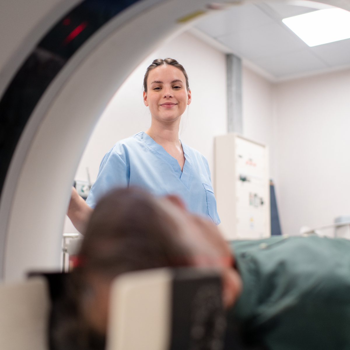
What Happens During the CT Scan?

  • Positioning: You will lie on a table that slides into the CT scanner. Depending on the area being scanned, you may be asked to hold still or hold your breath for a few seconds.
  • The scan: The CT scanner uses an X-ray tube that rotates around your body while taking images. The procedure is painless, but you may hear buzzing or clicking sounds from the machine.
  • Duration: The procedure typically takes around 5 to 20 minutes, though it may vary depending on the complexity of the scan.

After the CT Scan

  • If you had contrast dye, you might be asked to drink fluids to help flush it out of your system.

Risks and Considerations

  • Radiation exposure: CT scans involve exposure to X-rays, but the benefits of the scan typically outweigh the risks. The amount of radiation used is generally low, but pregnant women should avoid CT scans to prevent harm to the baby.
  • Contrast dye reactions: Some people may experience mild reactions to contrast dye, such as a rash or nausea. In rare cases, more serious reactions can occur, so inform your healthcare provider if you have a history of allergies or kidney problems.

Results and Follow-Up

  • After the CT scan, a radiologist will examine the images and send a report to your doctor.

What is CT Coronary Angiography?
It is a non-invasive examination that uses computed tomography (CT) to look at the blood vessels that supply the heart with blood (coronary arteries), the heart musculature, the chambers and function of the heart. This examination can detect blockages, known as plaques that are soft or calcified in nature and provide information about their location and the severity of the blockage to your doctor for necessary treatment. Iodinated contrast medium is used to highlight the blood vessels.

Why is CT Coronary Angiography Done?
Your doctor may recommend a CTCA if you are at risk for coronary artery disease (CAD) or have symptoms like chest pain, shortness of breath, or irregular heartbeats. It can help evaluate:

  • Blockages or narrowing of the coronary arteries
  • The presence of plaque buildup in the arteries
  • Abnormalities in the coronary anatomy

How should I prepare for the examination?

In CT coronary angiography, a slower heart rate makes it possible for the scanner to capture high-quality images of your blood vessels.

Please:

  • arrive early to your appointment to allow time for your heart rate to settle.
  • do not drink caffeinated drinks or smoke 24-hours prior to the exam
  • do not exercise on the day of the examination.
  • stop erectile dysfunction medication 3-days prior to the exam.

How is the Test Performed?

Preparation:

  • You will be asked to change into a gown. ECG leads (three dots) will be placed onto the chest to allow monitoring of the heart rate during the examination.
  • A small IV catheter will be placed into a vein in your arm.
  • A spray of GTN (angina medication) under the tongue will be given to dilate the heart arteries.

Procedure:

  • You will lie down on a table that slides into the CT scanner.
  • Contrast dye (usually iodine-based) will be injected into the catheter. You may feel some warmth or a flushed sensation from the contrast dye injection.
  • The CT scanner will rotate around you, taking multiple images to create detailed 3D pictures of your coronary arteries.
  • You will be asked to hold your breath for short periods (10-seconds) during the exam.
  • The test will take about 20-minutes.

What to Expect After the Test:

  • The results of the CTCA will be reviewed by a radiologist and discussed with your doctor, usually within a few days.
  • If you have received a contrast dye, be sure to drink plenty of fluids afterward to help clear it from your body.

Risks and Considerations:

  • Radiation Exposure: Although CTCA involves exposure to X-rays, the amount of radiation is relatively low. Your doctor will weigh the benefits and risks before recommending the test.
  • Allergic Reaction to Contrast Dye: While rare, some people may experience an allergic reaction to the dye. Be sure to inform your doctor if you have any known allergies or kidney problems.

What is a CT Arthrogram?
A CT arthrogram is an excellent way of imaging the internal structure of joints. An injected contrast dye allows for cartilage, ligaments and joint lining to be highlighted. Structures around the joint (tendons, muscles and bones) are also imaged.

Why Is a CT Arthrogram Needed?
This procedure is typically recommended to:

  • Diagnose joint problems, such as tears in cartilage, ligaments or labral tears.

Preparing for the Procedure

  • Medications: Please let us know if you are taking any blood thinning medication (warfarin, dabigatran, apixaban, aspirin or clopidogrel). You may need to have a blood test before the procedure and have your medication adjusted by your Doctor.
  • Allergies: Notify us if you have any allergies, especially to contrast dyes or iodine.
  • Pregnancy: If you are pregnant or suspect you might be, inform your healthcare provider. CT scans involve radiation, which may not be recommended during pregnancy.

What to Expect During the Procedure

  1. Positioning: Your will be asked to wear a gown. A radiologist will explain the procedure to you. You will be positioned on the CT scan table, and the area of the joint being examined will be cleaned and sterilized.
  2. Injection of Contrast Dye: A needle will be inserted into the joint to inject an iodine contrast dye, which may cause a sensation of fullness or pressure in the joint.
  3. CT Scan: After the injection, you will be asked to lie still while the CT scanner takes images of the joint. You may be asked to change positions during the scan to get different views.
  4. Duration: The procedure typically lasts 1 hour, including preparation time.

After the Procedure

  1. Post-Procedure Care: Once the procedure is complete, you may experience mild discomfort, swelling, or bruising at the injection site. These should resolve within hours. If pain persists beyond 24 hours please contact your doctor.
  2. Follow-up: Please book in to see your referring doctor to discuss the findings of the CT arthrogram.

Possible Risks and Complications
While a CT arthrogram is generally safe, there are some risks, including:

  • Radiation: You will receive a small dose of x-ray radiation.
  • Infection: Though rare, there is a small risk of infection at the injection site.
  • Allergic reaction: Some patients may have an allergic reaction to the contrast dye.
  • Bleeding: There is a very small risk of bleeding at the injection site.
  • Discomfort: Some discomfort or soreness at the injection site is common and should subside within a few days.

If you experience severe pain, swelling, redness, or fever after the procedure, contact your doctor immediately.

Patient Information Sheet:

A CT Calcium Score (also known as a coronary artery calcium (CAC) score) is a non-invasive imaging test that uses a computed tomography (CT) scan to detect the presence of calcium in the coronary arteries. The presence of calcium is an indicator of plaque buildup in the arteries, which can increase the risk of heart disease, heart attacks, or other cardiovascular problems.

The CT calcium score helps identify individuals at risk for coronary artery disease (CAD) even before symptoms appear. This early detection enables preventative treatment to be commenced before serious heart conditions develop.

Why is a CT Calcium Score Performed?

A CT Calcium Score is used to:

  • Assess risk for coronary artery disease (CAD), particularly in individuals who may not yet have symptoms (e.g., chest pain or shortness of breath).
  • Identify early-stage heart disease in people who may not yet show signs or symptoms.
  • Help determine the appropriate treatment and lifestyle changes, including medications, to reduce heart disease risk.
  • Monitor heart disease progression in individuals with known risk factors or pre-existing conditions (e.g., diabetes, hypertension, family history of heart disease).

Who Should Consider a CT Calcium Score?

Your doctor may recommend a CT Calcium Score if you are:

  • At intermediate risk for coronary artery disease and between the ages of 40–75.
  • A smoker, diabetic, or have high cholesterol, high blood pressure, or a family history of heart disease.
  • Concerned about your heart health, but do not yet show symptoms.
  • Trying to assess the risk of heart disease in a way that may guide decisions about treatment options, such as statins.

How is the Test Done?

  • The procedure is quick, non-invasive, and usually takes only 5-10 minutes.
  • You will be asked to lie down on a table that slides into the CT scanner.
  • Electrode stickers will be placed on your chest to monitor your heart’s electrical activity during the scan.
  • You may be asked to hold your breath briefly while the scan is performed to reduce motion and ensure clear images.
  • The CT scanner will produce detailed images of your heart, focusing on the coronary arteries.

What Does the CT Calcium Score Mean?

The calcium score reflects the amount of calcified plaque in your coronary arteries. The higher the score, the greater the amount of plaque and the higher the risk for heart disease. The results are generally classified as follows:

  • 0: No calcium detected – Low risk of heart disease.
  • 1–10: Minimal amount of calcium – Low risk.
  • 11–100: Mild amount of calcium – Moderate risk.
  • 101–400: Moderate amount of calcium – Increased risk.
  • Over 400: High amount of calcium – High risk.

Your score will help your doctor make decisions about treatment, lifestyle changes, or whether further testing is necessary.

Benefits of the CT Calcium Score

  • Non-invasive: No surgery or needles are required.
  • Quick and accurate: Provides detailed and clear images to assess plaque buildup.
  • Early detection: Helps identify individuals at risk of heart disease before symptoms arise.
  • Guides treatment: Helps your doctor decide whether you need medication or lifestyle changes.

Risks and Limitations

  • Radiation exposure: A CT scan does involve a small amount of radiation, but the risk is generally considered low. Your doctor will assess whether the benefits outweigh the risks.
  • Not suitable for everyone: This test may not be recommended for individuals with existing heart disease or those at very low risk for cardiovascular issues.
  • Limited to detecting calcium: The test does not show soft plaque (non-calcified) which may also pose a risk for heart attack.

What to Expect After the Test

  • Please book a follow up appointment with your doctor to discuss your results.

The test may be part of a larger strategy to help reduce your risk for heart disease, including lifestyle changes (e.g., diet, exercise), medications, or further tests.

A CT-guided biopsy is a medical procedure where a tissue sample is taken from a specific area inside your body using a CT (computed tomography) scan for guidance. The tissue sample is sent to a pathology lab for diagnosis.

Why is it done?

A CT-guided biopsy is used to obtain tissue from an abnormal area inside the body. It helps doctors to:

  • Determine if disease is present.
  • Determine the exact type of disease.
  • Plan for treatment options.

Before the Procedure

  • Preparation: Please do not eat or drink for 4 hours before the procedure.
  • Medications: Please inform us if you are taking any blood-thinning medication such as Aspirin, Warfarin, Dabigatran or Clopidogrel. You may need to have a blood-clotting test
  • before the procedure and have your medication adjusted by your doctor.

During the Procedure

  • Positioning: You will be asked to change into a gown and lie on a table. A CT scan will be used to locate the area where the biopsy will be done.
  • Planning: A safe biopsy path is planned and the entry site marked. Your skin will be cleansed to create sterile conditions, and a local anaesthetic is then injected.
  • Needle Insertion: A thin, hollow needle will be inserted through your numbed skin. You may be asked to hold your breath for a few seconds during the imaging. It is important that you try to keep as still as possible during the
  • procedure.
  • Sample Collection: Once the needle is in place, small tissue samples will be collected for testing. You may hear the noise of the biopsy equipment. This is normal.
  • Dressing: a dressing will be applied to the area.
  • Duration: The procedure typically takes 30-minutes.

After the Procedure

  • Observation: You will be monitored for 1-4 hours after the procedure to check for any immediate complications. You will need someone to drive you home and stay with you overnight.
  • Pain Management: Some soreness at the biopsy site is normal and paracetamol can help.
  • Dressing: you may remove your dressing one day after the procedure. You may bathe and shower as normal.
  • Activity Restrictions: Avoid physical exertion for 24 hours after the procedure.
  • Results: The pathology results will be sent to your doctor; this may take up to 7-days. You must return to your referring doctor to discuss the results.

Potential Risks and Complications

  • Radiation: You will receive a small dose of x-ray radiation. Please advise the radiographer if you are, or think you may be pregnant.
  • Bleeding: There may be some bleeding at the biopsy site. On rare occasions bleeding may be pronounced and require admission to hospital.
  • Infection: Any procedure involving a needle carries a small risk of infection.
  • Damage to surrounding organs or tissue: Though rare, there is a risk of injury to nearby organs or structures. We take every precaution to ensure your procedure is safe and sterile.
  • These risks must be weighed up against the risks of other options, like a surgical tissue sampling or not obtaining a diagnosis at all.

When to Contact Your Doctor

Call your doctor if you experience:

  • Severe pain at the biopsy site that doesn’t improve with pain medication.
  • Redness, swelling, or drainage at the site of the biopsy.
  • Difficulty breathing or chest pain (if the biopsy was near the lungs).
  • Fever or chills, which may be a sign of infection.

What is Virtual Colonoscopy?

Virtual colonoscopy is a non-invasive imaging technique that uses a CT scan (computerized tomography) to create detailed images of the colon (large intestine). This procedure allows doctors to check for abnormalities, such as polyps, tumours, or other conditions in the colon, without the need for traditional colonoscopy.

It is often used for screening purposes, particularly for patients who are at average risk for colon cancer or for those who cannot undergo a conventional colonoscopy.

Why is Virtual Colonoscopy Performed?

Virtual colonoscopy is typically done to:

  • Evaluate patients who are unable to undergo a traditional colonoscopy due to medical reasons
  • Screen for colorectal cancer (CRC) in asymptomatic individuals with average or moderate risk

What to Expect During the Procedure?

Preparation:

  • Bowel Prep: A clean colon is essential for the procedure to be effective. You will need to follow a special bowel prep that may include fasting, drinking a solution to clear your bowels, and avoiding certain foods or medications before the procedure.
  • Day of Procedure: You may be asked to arrive at the imaging centre 30 minutes to an hour before the procedure for final preparations.

Procedure:

  • You will lie on your back on the CT scan table. A small tube may be inserted into your rectum to gently inflate the colon with air. This helps to expand the colon, providing clearer images.
  • The CT scanner will take several images of your abdomen and pelvis from different angles. The procedure usually lasts 10 to 15 minutes.
  • You may be asked to change positions on the table to get images from different views.
  1. After the Procedure:

    • If air was used to inflate the colon, you might feel some discomfort or bloating. This usually resolves within a few hours.
    • Please book an appointment with your referring doctor to discuss your results.

Risks of Virtual Colonoscopy:

While virtual colonoscopy is generally safe, there are some risks to be aware of:

  • Radiation Exposure: As with all CT scans, there is some exposure to radiation. However, the amount used in virtual colonoscopy is minimal and considered safe for most people.
  • Inability to Perform Biopsy: If abnormalities such as polyps or tumors are found, a traditional colonoscopy may still be needed for biopsy or removal of the polyps.
  • Inconvenience or Discomfort: Some patients may experience discomfort from the air used to inflate the colon, or may feel bloated afterward.

Who Should Not Have Virtual Colonoscopy?

This procedure may not be suitable for patients who:

  • Are pregnant
  • Have had previous abdominal surgeries that might interfere with clear imaging.
  • Require colonoscopy. For high risk patients and certain types of bowel disease, such as active inflammatory bowel disease (IBD), traditional colonoscopy may be more appropriate.
  • Are unable to tolerate the bowel prep required for the procedure.

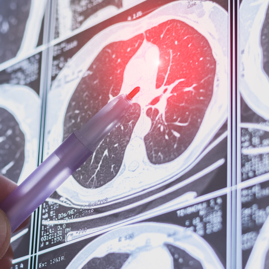
Australia’s New National Lung Cancer Screening Program is a screening tool designed to detect early signs of lung cancer, especially in high-risk individuals. Using advanced CT technology with lower radiation, it provides detailed images of the lungs to identify abnormalities such as tumours or lesions. This quick and non-invasive procedure can lead to earlier, more effective treatment

Patient Information Sheet:

The Australian government has introduced a new National Lung Cancer Screening Program aimed at detecting lung cancer at an early stage, when it is easier to treat. This program is for people who are at high risk of developing lung cancer and provides an opportunity for early detection, improving survival rates and outcomes.

Who is Eligible for Screening? The program is designed for people who:

  • Are aged 50 to 70 years.
  • Have a history of smoking 30 pack-years
    • A “pack year” is defined as smoking one pack of cigarettes per day for one year. For example, if you smoked one pack a day for 20 years, you would have 20 pack years.
  • Currently smoke or have quit smoking in the last 10-years

Please Note: People with symptoms of lung cancer, such as persistent cough, unexplained weight loss, or chest pain, should see their doctor immediately. This screening program is for people without symptoms but who are at high risk.

What is Lung Cancer Screening?

Lung cancer screening involves a test called a low-dose CT scan (LDCT), which takes detailed images of the lungs. This test can help detect early signs of lung cancer before symptoms appear, increasing the chances of successful treatment.

How Does the Screening Work?

  1. Referral from Your GP: If you are eligible, your GP will refer you for a low-dose CT scan. If you’re unsure whether you’re eligible, ask your GP.
  2. The Low-Dose CT Scan: The screening involves lying on a table while a CT scanner takes pictures of your lungs. The procedure is quick, painless, and usually takes about 10 minutes. You may need to hold your breath briefly during the scan.
  3. Results: After the scan, a specialist radiologist will review the images. If no concerning abnormalities are found, you will be informed, and the next screening appointment will be scheduled in 2 years. If anything suspicious is detected, further tests or consultations with a lung cancer specialist may be recommended.

Benefits of Lung Cancer Screening:

  • Early Detection: Screening can detect lung cancer at an early stage when it is more treatable.
  • Improved Outcomes: Early treatment significantly improves survival rates for lung cancer patients.

Possible Risks and Considerations:

  • False Positives: Sometimes the screening may suggest something abnormal, but further testing may show that it is not cancer. This can cause anxiety and unnecessary follow-up procedures.
  • False Negatives: In rare cases, the screening may miss some cancers, especially if they are in the early stages.
  • Radiation Exposure: The low-dose CT scan involves a small amount of radiation. However, the benefit of early cancer detection outweighs the risks associated with this radiation.

What Happens After Screening?

  • No Further Action: If your results are clear, you will be asked to return for another screening in two years.
  • Further Testing: If the scan shows something abnormal, your doctor may recommend additional tests, such as a biopsy, bronchoscopy, or PET scan, to determine whether you have lung cancer or another condition.

Important Reminders:

  • Don’t Skip Your Screening: If you are eligible for the program, regular screening is important for early detection.
  • Talk to Your Doctor: If you have any concerns or questions about the screening process, talk to your doctor. They can help guide you through the program and discuss whether it’s right for you.

Contact Information:

If you have questions about the National Lung Cancer Screening Program or want to learn more, you can contact the following:

  • Your General Practitioner (GP) for more details about your eligibility and the referral process.
  • The National Cancer Screening Program hotline: 1800 627 701
  • For further information, visit the official National Lung Cancer Screening Program website: https://www.health.gov.au/our-work/nlcsp

Are CTs Safe?

You will receive a small dose of x-ray radiation. Please advise the radiographer if you are, or think you may be pregnant.

After the Exam

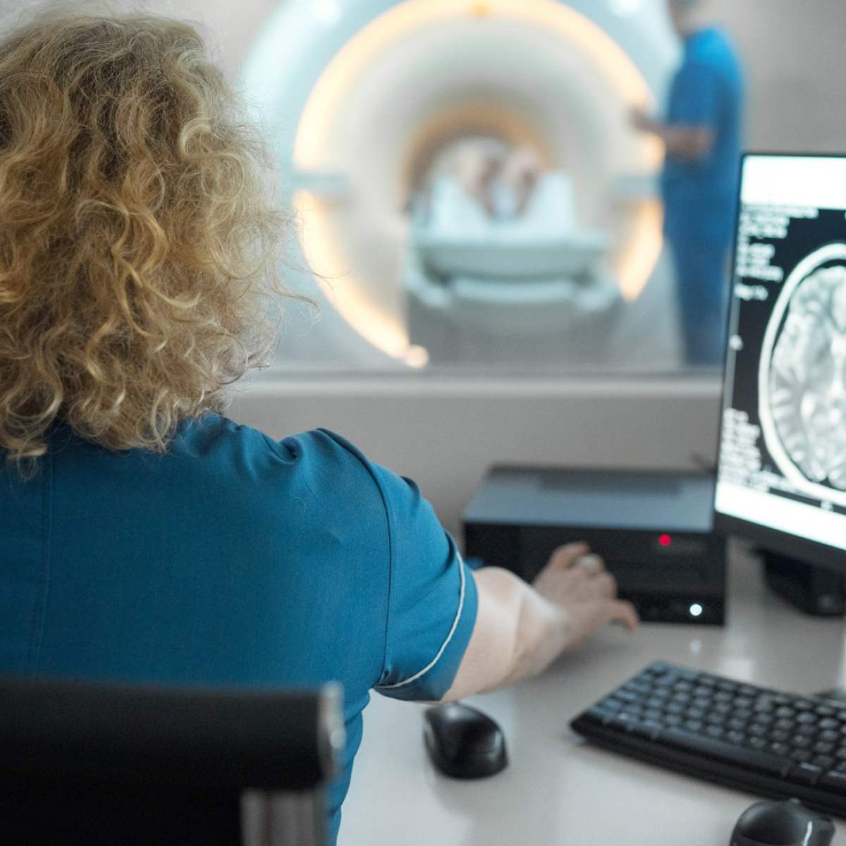
The CT images will be reviewed by a radiologist and sent to your doctor. Please book an appointment with your referring doctor to discuss the findings. Most results are available within a few days.

Key Features of Our Clinic

Our point of difference

All Referrals Welcome

In Australia, radiology referrals are not clinic-specific. You can use a referral made out to another clinic here.

Expert Radiologists

A study is only as good as the specialist reporting it. Our team are experienced and broadly knowledgeable radiologists with sub-specialised training.

Latest Low-Dose Technology

We have invested in the latest imaging technology to ensure our patients enjoy the safest clinical experience possible.

Fast Reporting to GPs & Specialists

Onsite Radiologists means rapid turn-around of reports, with written correspondence sent within 2–24hours.

Urgent & Emergency

Urgent appointments are always accommodated. Simply call reception.

Our Team

Meet Our Radiology Team

Dr Andrew Low

BEng, MSc, MBBS(Hons 1), FRCPC, FRANZCR

Radiologist

Dr James Bewes

BScAdv, MMedPhys, MBBS(Hons), FRANZCR

Radiologist

Questions

If you have any questions or concerns about the procedure, please feel free to contact our friendly staff.

Expert imaging. Clear answers. Compassionate care. Book your appointment today!

If you wish to make a booking or require urgent attention, please get in touch with our friendly team to make a booking.

Please Note

We are NOT open to the public yet as our clinic is still under construction. Our official opening date is scheduled to be late February 2026.