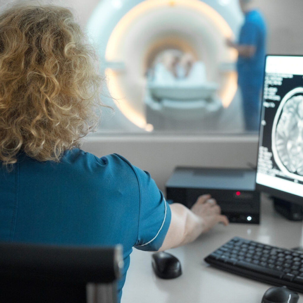
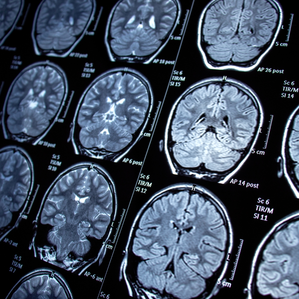
An MRI scan is a non-invasive imaging technique that provides detailed, high-resolution images of the body’s internal structures, including the brain, spine, joints, and soft tissues.
It helps diagnose conditions like musculoskeletal injuries, infections, tumours, and neurological disorders.
Using powerful magnets and radio waves, MRI offers clear, accurate results without radiation.
A MRI scan is a medical imaging technique used to create detailed pictures of the inside of your body. MRI uses strong magnets, radio waves, and a computer to generate images of your organs, soft tissues, and bones.
What is an MRI?
A general Magnetic Resonance Imaging (MRI) scan is a non-invasive imaging technique that provides detailed, high-resolution images of the body’s internal structures, including the brain, spine, joints, and soft tissues.
It helps diagnose conditions like injuries, infections, tumours, and neurological disorders. Using powerful magnets and radio waves, MRI offers clear, accurate results without radiation.
Preparing for Your MRI
Safety Questionnaire:
On arrival you will be asked to complete a safety questionnaire. This is to ensure it is safe for you to have the scan. You can also complete this questionnaire online prior to your appointment here.
It is not safe for certain people with certain metallic implants to have an MRI due to the strong magnetic field. Any implanted medical devices such as pacemakers, insulin pumps, brain aneurysm clips, ear implants or metallic foreign bodies may exclude a patient from having an MRI.
An MRI may be unsuited to those that suffer from any of the following:
Clothing:
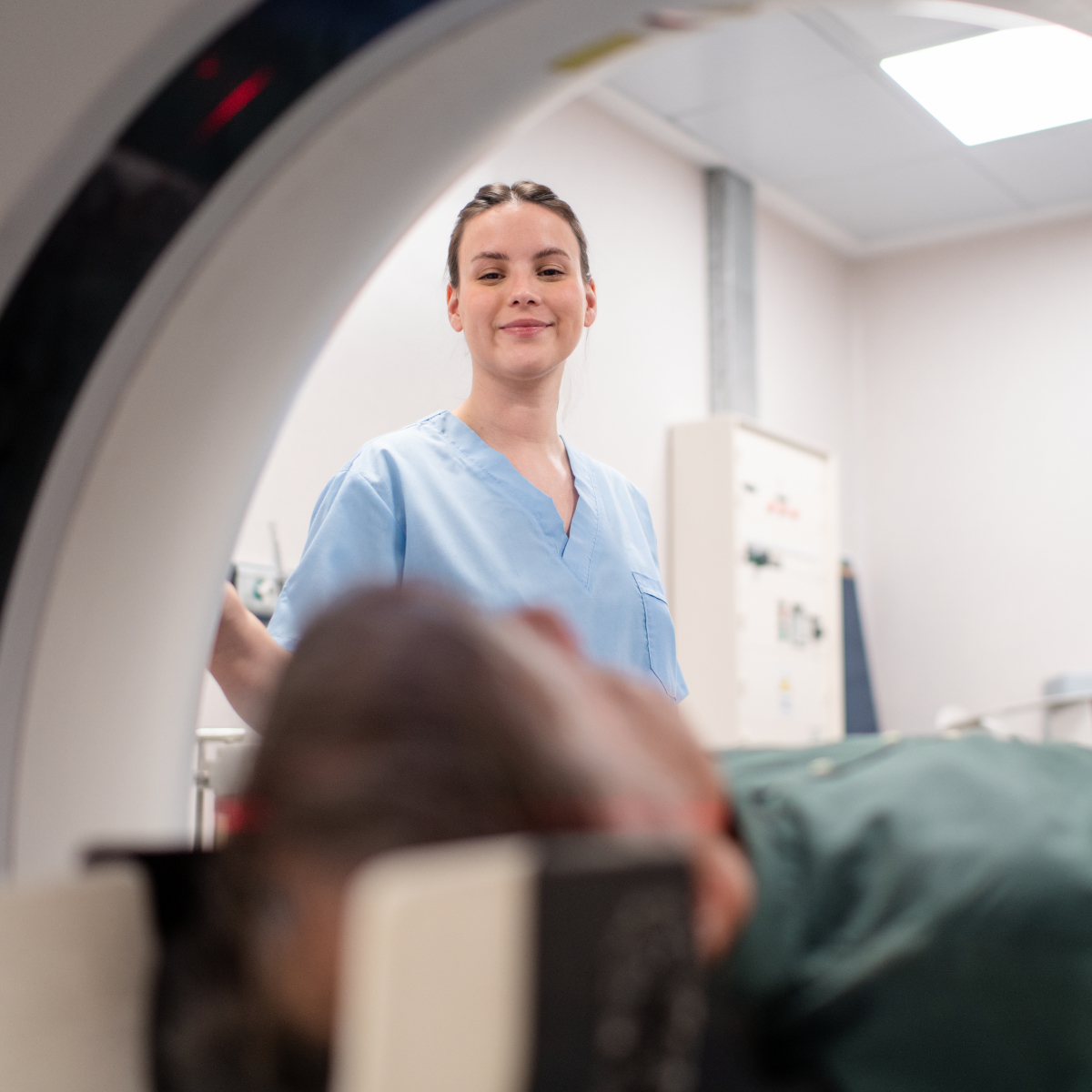
You will be asked to change into a gown and remove all jewellery, hairpins, and any metal objects before entering the MRI room. This includes credit cards, watches, and hearing aids. You will be asked to leave your belongings in a secure area. Underpants and socks may be worn.
What Happens During the MRI?
How Long Does the MRI Take?
An MRI typically lasts 20 to 45 minutes, depending on the area being scanned. Some scans may take longer, especially if contrast dye is used.
After the MRI
Is the MRI Safe?
MRI is generally very safe. It doesn’t use radiation, unlike X-rays or CT scans. However, the strong magnets can interact with certain metals, which is why it’s important to disclose any medical implants or devices you may have. Pregnant women should avoid MRI in the first trimester unless absolutely necessary.
What is a Prostate MRI?
Prostate MRI is a non-invasive imaging procedure used to assess the prostate gland for conditions such as cancer, benign enlargement, or inflammation. It provides detailed images to help guide diagnosis and treatment planning. Using advanced MRI technology, this procedure offers high-resolution images without radiation, ensuring accurate results.
It helps doctors diagnose, evaluate, and monitor prostate conditions, including prostate cancer.
Why Might I Need a Prostate MRI?
The most common cancer to affect males is prostate cancer. MRI can be used to:
Preparation for the Procedure:
Safety Questionnaire:
On arrival you will be asked to complete a safety questionnaire. This is to ensure it is safe for you to have the scan. You can also complete this questionnaire online prior to your appointment here.
It is not safe for people with certain metallic implants to have an MRI due to the strong magnetic field. Any implanted medical devices such as pacemakers, insulin pumps, brain aneurysm clips, ear implants or metallic foreign bodies may exclude a patient from having an MRI.
Clothing:
You will be asked to change into a gown and remove all jewellery, hairpins, and any metal objects before entering the MRI room. This includes credit cards, watches, and hearing aids. You will be asked to leave your belongings in a secure area. Underpants and socks may be worn.
Contrast Dye:
A contrast dye and a small bowel muscle relaxant will be administered via an intravenous cannula inserted into you. This stops the bowel moving for 15-minutes during the exam and allows visualisation of the prostate.
What to Expect During the Procedure:
After the MRI:
Is the prostate MRI Safe?
MRI is generally very safe. It doesn’t use radiation, unlike X-rays or CT scans. However, the strong magnets can interact with certain metals, which is why it’s important to disclose any medical implants or devices you may have.
Contrast Dye Reactions:
Rarely, people may have an allergic reaction to the contrast dye. Inform your doctor if you have any known allergies, particularly to iodine or shellfish.
What is a Cardiac MRI?
Cardiac MRI is a non-invasive imaging technique used to assess the heart’s structure and function. It provides detailed images of the heart muscle, blood vessels, and valves. It is the best test (gold standard) for many cardiac conditions.
Why Might I Need a Cardiac MRI?
Your doctor may request a Cardiac MRI to:
How Do I Prepare?
Before the Scan:
What Happens During the Scan?
After the Scan
Risks and Safety
What is a Rectal MRI?
An MRI of the rectum is a specialised imaging procedure used to evaluate the function and structure of the rectum and pelvic floor. It is commonly used to diagnose conditions such as rectal prolapse, incontinence, or pelvic floor dysfunction. This non-invasive procedure uses MRI technology to provide detailed, high-resolution images, aiding in accurate diagnosis and treatment planning.
How Do I Prepare?
Before the Scan:
What Happens During the Scan?
On Arrival:
In the MRI Scanner:
After the MRI
Important Notes
Please inform us before the scan if you:
What Is A Pelvic MRI?
A pelvic MRI acquires detailed images of organs and structures in the pelvic area. The test is safe, painless and does not use radiation. A pelvic MRI can be used to evaluate the uterus, ovaries and their surrounding tissues and structures. This non-invasive procedure uses MRI technology to provide detailed, high-resolution images, aiding in accurate diagnosis and treatment planning.
Safety Questionnaire:
On arrival you will be asked to complete a safety questionnaire. This is to ensure it is safe for you to have the scan. You can also complete this questionnaire online prior to your appointment here.
It is not safe for some people with certain metallic implants in their bodies to have an MRI due to the strong magnetic field. Any implanted medical devices such as pacemakers, insulin pumps, brain aneurysm clips, ear implants or metallic foreign bodies may exclude a patient from having an MRI.
Clothing:
You will be asked to change into a gown and remove all jewellery, hairpins, and any metal objects before entering the MRI room. This includes credit cards, watches, and hearing aids. You will be asked to leave your belongings in a secure area. Underpants and socks may be worn.
What to Expect During the Procedure
After the MRI
If you had contrast dye, you may be advised to drink plenty of fluids to help flush it from your system.
Is the MRI Safe?
MRI is generally very safe. It doesn’t use radiation, unlike X-rays or CT scans. However, the strong magnets can interact with certain metals, which is why it’s important to disclose any medical implants or devices you may have.
Contrast Dye Reactions:
Rarely, people may have an allergic reaction to the contrast dye. Inform your doctor if you have any known allergies, particularly to iodine or shellfish.
What is an MRI Arthrogram?
An MRI arthrogram is a special type of MRI (Magnetic Resonance Imaging) that is used to take detailed pictures of the inside of a joint. Unlike a regular MRI, an MRI arthrogram involves injecting a contrast dye into the joint before the scan. This dye helps to highlight areas within the joint and allows for a clearer and more detailed view of the cartilage, ligaments, tendons, and other structures.
Common Joints for MRI Arthrograms:
Why is an MRI Arthrogram Performed?
An MRI arthrogram is typically recommended when:
Before the Procedure
Preparation:
Clothing:
During the Procedure
Step 1: Injection of Contrast Dye
Step 2: MRI Scan
After the Procedure
When to Contact Your Doctor
Possible Risks and Complications
Although MRI arthrograms are generally safe, there are some risks to be aware of:
If you experience any severe reactions, it is important to contact your healthcare provider immediately.
What is an MRI Enterography?
An MRI enterogram is a specialised imaging test that takes detailed images of your small intestine. It helps doctors visualise inflammation, blockages or other problems in the intestines without using radiation.
How Do I Prepare?
What Happens During the Scan?
On Arrival:
In the MRI Scanner:
After the MRI Enterogram
An MRI is generally very safe. Unlike X-rays or CT scans, MRI scans do not use radiation. However, the strong magnets can interact with certain metals, which is why it’s important to disclose any medical implants or devices you may have. Pregnant women should avoid MRI in the first trimester unless absolutely necessary.
In Australia, radiology referrals are not clinic-specific. You can use a referral made out to another clinic here.
An imaging study is only as good as the specialist reporting it. Our team are highly-experienced radiologists, with sub-specialised areas of expertise.
We have invested in the latest low-dose imaging technology, which offer enhanced clarity, to ensure our patients enjoy the safest clinical experience possible.
Onsite Radiologists means rapid turn-around of reports, with results sent back to your referrer within 24-hours, or the next working day.
Clinically urgent appointments are always accommodated. Please call reception for further assistance.
If you have any questions or concerns about MRIs, please feel free to contact our friendly staff.
If you wish to make a booking or require urgent attention, please get in touch with our friendly team to make a booking.


Welcome to our brand new clinic!
The majority of our services have same-day or next-day availability (excluding MRI and more complex examinations).
Our Services:
Please phone or email our team to make a booking. Online bookings also coming soon!
All referrals accepted.