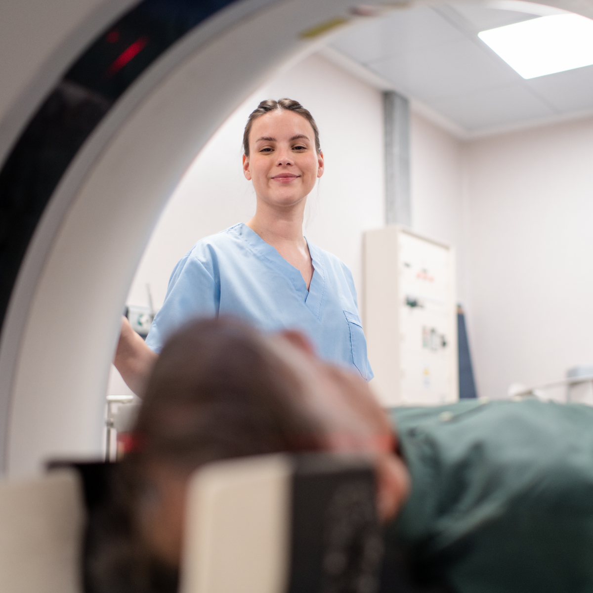
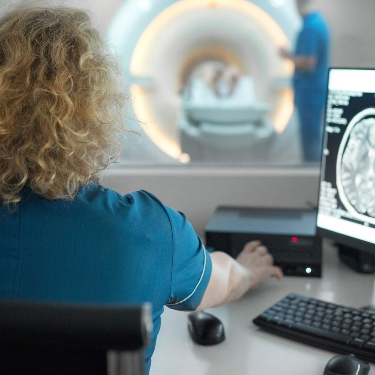
Ultrasound and CT guided interventional procedures encompass a wide range of injections, biopsies and other therapeutic and diagnostic procedures which assist your referring practitioner in managing a condition or creating a treatment plan for a diagnosed issue.
Our technical staff and radiologists all provide the highest level of care and expertise when undergoing these procedures.
Please see below for information for some of the most commonly referred services that we provide.

What is an Ultrasound Guided Injection?
An ultrasound guided joint injection is a non-surgical treatment used to relieve pain and allow improved mobility in the joint. It may involve injection of a corticosteroid in conjunction with a local anaesthetic to relieve pain and inflammation. In patients with joint injury or osteoarthritis, particularly in the knee, hyaluronic acid can be injected into the joint to restore lubrication and function. Guided by imaging techniques for precision, this treatment helps reduce pain and improve function.
Before your appointment
During the Procedure
After the Procedure
Contact your doctor if you experience:
Potential Risks (uncommon)
What is an Ultrasound Guided Biopsy?
An ultrasound-guided biopsy is a minimally invasive procedure used to obtain tissue samples. Real-time ultrasound imaging is used to help guide a needle to an area where a tissue sample is needed. The needle is then inserted into the tissue, and a small sample is taken for testing in a laboratory.
The procedure ensures precise needle placement, allowing for accurate tissue sampling. This diagnostic tool helps identify conditions such as cancer, cysts, or infections.
By obtaining a tissue sample, the doctor can determine whether a condition is benign or malignant and recommend the best course of treatment.
How is the Procedure Done?
Preparation:
Ultrasound Imaging:
Biopsy Procedure:
After the Procedure:
What to Expect After the Procedure?
Potential Risks and Complications?
While ultrasound-guided biopsies are generally safe, there are some potential risks, including:
If you experience any unusual symptoms after the biopsy, such as excessive pain, swelling, redness, fever, or if you notice blood in your urine, stool, or vomit, contact your doctor immediately.
Frequently Asked Questions
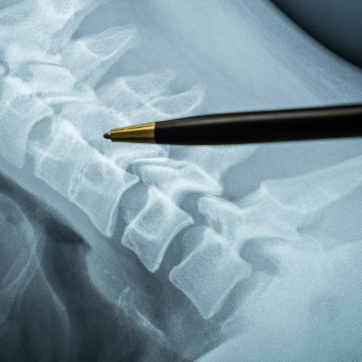
What is a CT Guided Facet Joint Injection?
A CT-guided facet joint injection is a procedure in which anti-inflammatory medication is injected into one or more facet joints in the spine. The injection is performed using CT (computed tomography) imaging to accurately guide the needle to the exact location. Facet joints are small joints between each vertebra in the spine. They help with movement and stability but can become painful due to arthritis, injury, or inflammation. Injecting steroid and local anaesthetic into a facet joint may confirm whether the facet joints are the source of your pain and in turn relieve that pain.
Before Your Procedure
Please inform our staff if you:
During Your Procedure
After your procedure
Aftercare Instructions
Contact your provider right away if you experience:
Possible Risks
What is a Nerve Root Injection?
A nerve root injection, (also referred to as periradicular injection), is a procedure used to treat nerve pain which originates in the back. Using CT guidance, a corticosteroid and anaesthetic are injected around the affected nerve root. This reduces inflammation around the nerve root, thereby relieving neuropathic (nerve) pain radiating to the buttock or leg. This minimally invasive treatment can determine if a specific nerve is causing your pain and if so, can provide effective, long-lasting relief.
Before Your Procedure
Please inform our staff if you:
During Your Procedure
After Your Procedure
Aftercare instructions
Contact your doctor right away if you experience:
Possible Risks
What is an Epidural Injection?
An epidural injection is a minimally invasive procedure used to relieve pain and inflammation in the spine. Guided by imaging techniques, such as fluoroscopy or CT, the injection delivers medication directly into the epidural space around the spinal cord. It is commonly used to treat conditions like sciatica, herniated discs, and spinal stenosis.
Why is an Epidural Injection Done?
A common cause of sciatic pain is inflammation in response to nerve irritation caused by nerve entrapment or spinal disc protrusion. An injection of anti-inflammatory medication (steroids) into the epidural space can reduce this inflammation and decrease pain.
How is the Procedure Performed?
What to Expect After the Procedure
Effects From the Treatment
The local anaesthetic may give temporary relief from pain for up to four hours. Paracetamol may be taken if you experience discomfort. The steroid can take up to a week to reach its maximum effect. Relief from symptoms does vary between patients, both in time and scale. Some people do not receive pain relief from the procedure if inflammation is not the main cause for their pain. Please keep in mind this is generally not a failure of the procedure. This information is useful for your doctor as it indicates other causes of pain that may need to be considered.
What Are the Risks?
What is a CT Guided Biopsy?
A CT-guided biopsy is a medical procedure where a tissue sample is taken from a specific area inside your body using a CT (computed tomography) scan for guidance. The tissue sample is sent to a pathology lab for diagnosis.
It is used to obtain tissue from an abnormal area inside the body. It helps doctors to:
Before the Procedure
During the Procedure
After the Procedure
Potential Risks and Complications
Call your doctor if you experience:
What is Shoulder Hydrodilatation?
Shoulder hydrodilatation is a minimally invasive procedure used to treat frozen shoulder (adhesive capsulitis) by injecting a sterile fluid mixture into the shoulder joint. This helps to stretch the joint capsule, improving range of motion and reducing pain. Guided by imaging techniques for precision, the procedure provides effective relief for patients with limited shoulder mobility.
Before your procedure
Tell our staff if you:
Preparation
During Your Procedure
After the Procedure
What are the benefits?
Are there any risks?
Most people tolerate the procedure well. Side effects are rare but may include:
When to seek medical help?
Call your doctor if you experience:
What happens next?
Yes, interventional radiology is considered very safe as procedures are minimally invasive, there are only a small amount of risks and complications are rare.
In Australia, radiology referrals are not clinic-specific. You can use a referral made out to another clinic here.
An imaging study is only as good as the specialist reporting it. Our team are highly-experienced radiologists, with sub-specialised areas of expertise.
We have invested in the latest low-dose imaging technology, which offer enhanced clarity, to ensure our patients enjoy the safest clinical experience possible.
Onsite Radiologists means rapid turn-around of reports, with results sent back to your referrer within 24-hours, or the next working day.
Clinically urgent appointments are always accommodated. Please call reception for further assistance.
If you have any questions or concerns about the procedure, please feel free to contact our friendly staff.
If you wish to make a booking or require urgent attention, please get in touch with our friendly team to make a booking.

Welcome to our brand new clinic!
The majority of our services have same-day or next-day availability (excluding MRI and more complex examinations).
Our Services:
Please phone or email our team to make a booking. Online bookings also coming soon!
All referrals accepted.