Most ultrasounds are painless and non-invasive, though some may involve slight pressure or, in the case of certain pelvic exams, an internal probe. Depending on the type of scan, you might be asked to fast beforehand, drink water to fill your bladder, or wear loose clothing. Most examinations usually take between 15 and 45 minutes. Overall, ultrasound is a safe and non-invasive imaging method with no radiation exposure.
Gibraltar Radiology perform a wide range of general, musculoskeletal, arterial, vascular and obstetric ultrasounds.
Please see below for information on some of the most commonly referred services that we provide.
What is a Musculoskeletal Ultrasound Scan?
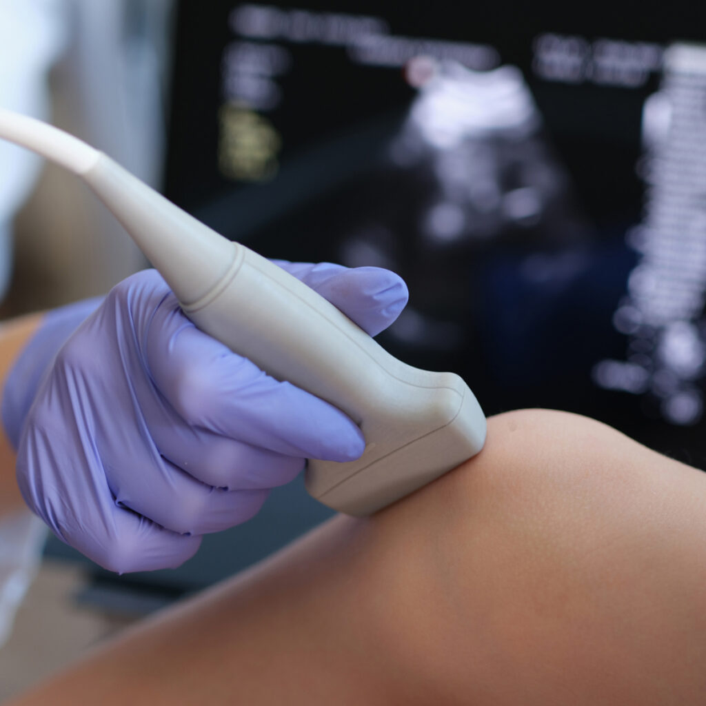
A musculoskeletal ultrasound is a non-invasive imaging technique that can be used to provide detailed images of muscles, tendons and ligaments throughout the body. Ultrasound scans can assess and diagnose injuries, inflammation as well as causes of acute or chronic musculoskeletal pain. An x-ray of the region may be recommended. This provides assessment of bony structures not seen on ultrasound.
This safe, radiation-free procedure provides real-time images, aiding in accurate diagnosis and treatment planning.
Why Might I Need This Type of Scan?
Your doctor may recommend a musculoskeletal ultrasound if you have suspected:
What Happens During the Ultrasound?
Is Musculoskeletal Ultrasound Safe?
Yes. There are no known risks associated with ultrasound, making it a preferred method for imaging, especially for pregnant women, and children.
After the Exam
The ultrasound images will be reviewed by a radiologist and the results sent to your doctor. Please book an appointment with your referring doctor to discuss the findings. Results will be available within 24 hours or the next working day.
What is an Upper-Abdominal Ultrasound?
Ultrasound scans can visualise your liver, gallbladder, spleen, pancreas and kidneys. It helps diagnose conditions like kidney stones, liver disease, or gallstones. Not all causes of abdominal pain can be identified with ultrasound, but because this test is safe and free from radiation, it will often be used as an initial investigation.
Why Might I Need This Type of Scan?
Your doctor may recommend an upper abdominal ultrasound if you are experiencing symptoms like:
How Should I Prepare?
For an upper abdomen ultrasound, preparation is typically required:
What Happens During the Ultrasound?
Is the Ultrasound Safe?
Yes, an ultrasound is very safe. It uses sound waves instead of radiation. It is a non-invasive and painless procedure.
After the Ultrasound
The ultrasound images will be reviewed by a radiologist and the results sent to your doctor. Please book an appointment with your referring doctor to discuss the findings. Results will be available within 24 hours or the next working day.
What is Liver Elastography?
Liver elastography is a specialised, non-invasive imaging technique used to assess liver stiffness, helping to detect conditions like fibrosis, cirrhosis, and fatty liver disease. Using ultrasound technology, this examination measures tissue elasticity, providing valuable insights into liver health without the need for a biopsy. Safe and painless, elastography aids in early diagnosis and monitoring. This test is used to assess the degree of liver fibrosis (scarring) caused by conditions such as:
Why Might I Need this Type of Scan?
Your healthcare provider may recommend liver elastography to:
Unlike a liver biopsy, elastography is a non-invasive, quick, and painless procedure.
How Should I Prepare?
What Happens During the Ultrasound?
Is Liver Elastography Safe?
Yes, liver elastography is safe, with no known side effects. Unlike a biopsy, there is no risk of bleeding or infection.
Who Should Not Have Liver Elastography?
While liver elastography is safe for most people, it may not be suitable if:
Please inform your healthcare provider if any of these conditions apply to you.
After the Ultrasound
The ultrasound images and measurements will be reviewed by our radiologist and the results sent to your doctor, who will further interpret the results. Please book an appointment with your referring doctor to discuss the findings. Results will be available within 24 hours or the next working day.
What is a Renal and Urinary Bladder Ultrasound?
Renal and urinary bladder ultrasounds are a non-invasive imaging technique used to assess the kidneys and bladder. It helps diagnose conditions like kidney stones, cysts and tumours, infections, hydronephrosis and bladder abnormalities.
Using high-frequency sound waves, this safe procedure provides clear, detailed images for accurate diagnosis and treatment planning.
Why Might I Need This Type of Scan?
Your doctor may recommend an upper abdominal ultrasound if you are experiencing symptoms like:
How Should I Prepare?
For a renal and urinary bladder ultrasound, preparation is typically required:
What Happens During the Ultrasound?
Is the Ultrasound Safe?
Yes, an ultrasound is very safe. It uses sound waves instead of radiation. It is a non-invasive and painless procedure.
After the Ultrasound
The ultrasound images will be reviewed by a radiologist and the results sent to your doctor. Please book an appointment with your referring doctor to discuss the findings. Results will be available within 24 hours or the next working day.
What is a Pelvic Ultrasound?
Pelvis and gynaecological ultrasounds are imaging tests that use sound waves to create images of the organs and structures in your pelvic area, including the uterus & ovaries. It helps your doctor diagnose and monitor various conditions such as endometriosis, cysts, fibroids or uterine abnormalities.
Why Might I Need This Type of Scan?
Your doctor may recommend a pelvic ultrasound to:
Booking Your Exam
Ultrasound can be performed at any time of the menstrual cycle. However, interpreting the appearance of the ovaries and endometrium (lining of the uterus) is ideal immediately after your period. Please always follow any specific instructions from your referring practitioner with regards to examination timing.
How Should I Prepare?
What Happens During the Ultrasound?
What is a Transvaginal Ultrasound?
A transvaginal ultrasound provides the best views of the ovaries and uterus, and in most cases is recommended. For this, a thin probe is inserted into the vagina to provide closer images of your uterus and ovaries.
You may be asked to empty your bladder before this part of the exam.
Transvaginal scans are not performed on young girls, those who have not been sexually active or those who choose not to have this type of scan.
If this exam is not appropriate for you, please let the sonographer know.
Is The Ultrasound Safe?
Vaginal ultrasound probes are sterilised and covered by a disposable sheath for each patient. There are no known risks associated with ultrasound.
After the Exam
The ultrasound images will be reviewed by our radiologist and the results will be sent to your doctor. Please book in an appointment with your referring doctor to discuss the findings. Results will be available within 24 hours or the next working day.
What is an Early Pregnancy Ultrasound?
An early pregnancy ultrasound is a non-invasive imaging procedure that uses sound waves to create an image of your pregnancy in the womb. This ultrasound is typically performed in the first trimester, usually between 6 and 10 weeks of pregnancy, to help confirm and evaluate the early stages of pregnancy.
Why Might I Need This Type of Scan?
Your healthcare provider may recommend an early pregnancy ultrasound for several reasons, including:
How Should I Prepare?
You will be asked to have a full bladder. Please drink as close to 1L of water 1 hour before your examination and do not urinate until your examination is complete.
What Happens During the Ultrasound?
Is the Ultrasound Safe?
Ultrasound is a very safe procedure, with no known risks to you or your baby. It does not use radiation like X-rays and is widely regarded as a safe way to monitor the progress of a pregnancy.
After the Ultrasound
The images will be reviewed by our radiologist and the results sent to your doctor. Please book an appointment with your referring doctor to discuss the findings. Results will be available within 24hrs or the next working day.
What is an Obstetric Ultrasound?
An obstetric ultrasound is a non-invasive imaging procedure that uses sound waves to create pictures of your baby in the womb. It provides valuable information about your baby’s growth, development, and general health during pregnancy. Ultrasound is safe for both you and your baby and is a routine part of prenatal care.
Types of Obstetric Ultrasound Performed
12-16wk Early Morphology
An early morphology ultrasound, typically performed in the first trimester, ideally around 14 weeks, is used to assess the baby’s development, check for a heartbeat, and confirm the due date based on the size of the foetus.
Nuchal Translucency
The Nuchal Translucency (NT) scan is usually performed between 12-13 weeks. It measures the fluid at the back of your baby’s neck. The amount of fluid present is one of the markers used to assess the risk of certain chromosomal conditions, such as Down syndrome (trisomy 21), trisomy 18, and trisomy 13. A higher-than-normal amount of fluid may indicate an increased risk for chromosomal abnormalities, though many babies with a thicker NT fold are healthy. The NT scan is usually combined with blood tests to give a more complete risk assessment.
20-Week Morphology Ultrasound
The 20-week morphology ultrasound, also known as the anatomy scan, occurs in the second trimester and provides a detailed look at the baby’s organs, bones, and overall growth. This scan helps identify structural abnormalities such as problems with the heart, brain, spine, kidneys, or limbs. It also provides information on the location of the placenta and how much amniotic fluid is present. It can also be used to confirm the due date, and, if desired, reveal the baby’s gender. This ultrasound is important for monitoring pregnancy health and foetal development.
What Happens During the Ultrasound?
You’ll lie on your back, and gel will be applied to your belly. The technician will use a transducer to capture images of your baby. You may be able to see your baby’s features and movements on the screen, which can be a very exciting moment! The scan usually takes anywhere between 30-60 minutes, depending on what stage of pregnancy you are at.
How Should I Prepare?
Is There Any Risk to My Baby?
No, ultrasound is considered a safe procedure during pregnancy. It does not use radiation, and there is no evidence to suggest it poses a risk to you or your baby.
After the Ultrasound
The ultrasound images will be reviewed by our radiologist and the results will be sent to your doctor. Please book an appointment with your referring doctor to discuss the findings. Results will be available within 24 hours or the next working day.
Most of the time, the results will be normal, and no further action will be needed. If there are any concerns, your healthcare provider may suggest further testing or refer you to a specialist.
Yes. There are no known risks associated with ultrasound, making it a preferred method for imaging, especially for pregnant women, and children.
In Australia, radiology referrals are not clinic-specific. You can use a referral made out to another clinic here.
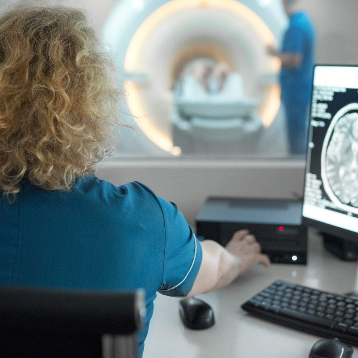
An imaging study is only as good as the specialist reporting it. Our team are highly-experienced radiologists, with sub-specialised areas of expertise.
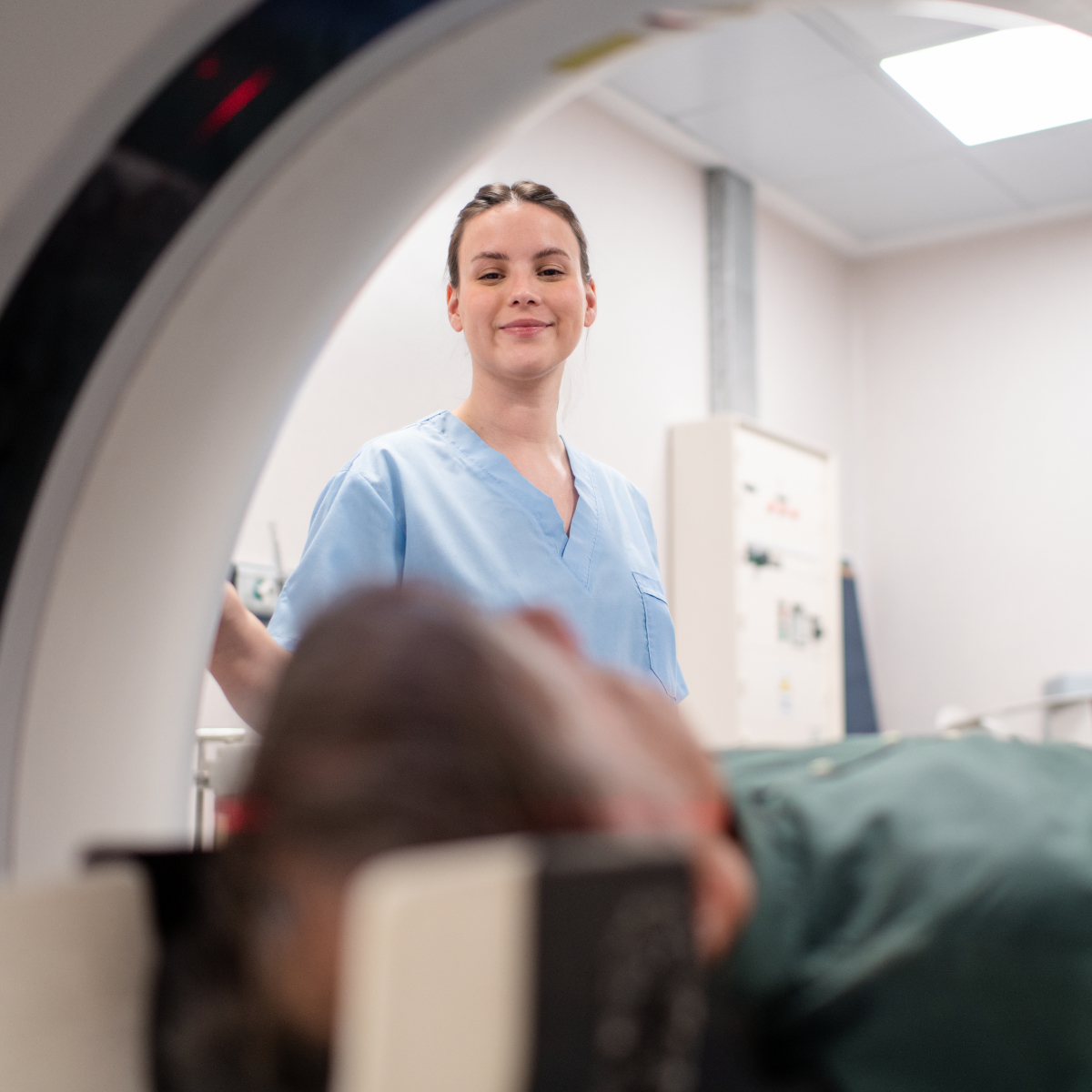
We have invested in the latest low-dose imaging technology, which offer enhanced clarity, to ensure our patients enjoy the safest clinical experience possible.
Onsite Radiologists means rapid turn-around of reports, with results sent back to your referrer within 24-hours, or the next working day.
Clinically urgent appointments are always accommodated. Please call reception for further assistance.
In Australia, radiology referrals are not clinic-specific. You can use a referral made out to another clinic here.
An imaging study is only as good as the specialist reporting it. Our team are highly-experienced radiologists, with sub-specialised areas of expertise.
We have invested in the latest low-dose imaging technology, which offer enhanced clarity, to ensure our patients enjoy the safest clinical experience possible.
Onsite Radiologists means rapid turn-around of reports, with results sent back to your referrer within 24-hours, or the next working day.
Clinically urgent appointments are always accommodated. Please call reception for further assistance.
If you have any questions or concerns about ultrasounds, please feel free to contact our friendly staff.
If you wish to make a booking or require urgent attention, please get in touch with our friendly team to make a booking.